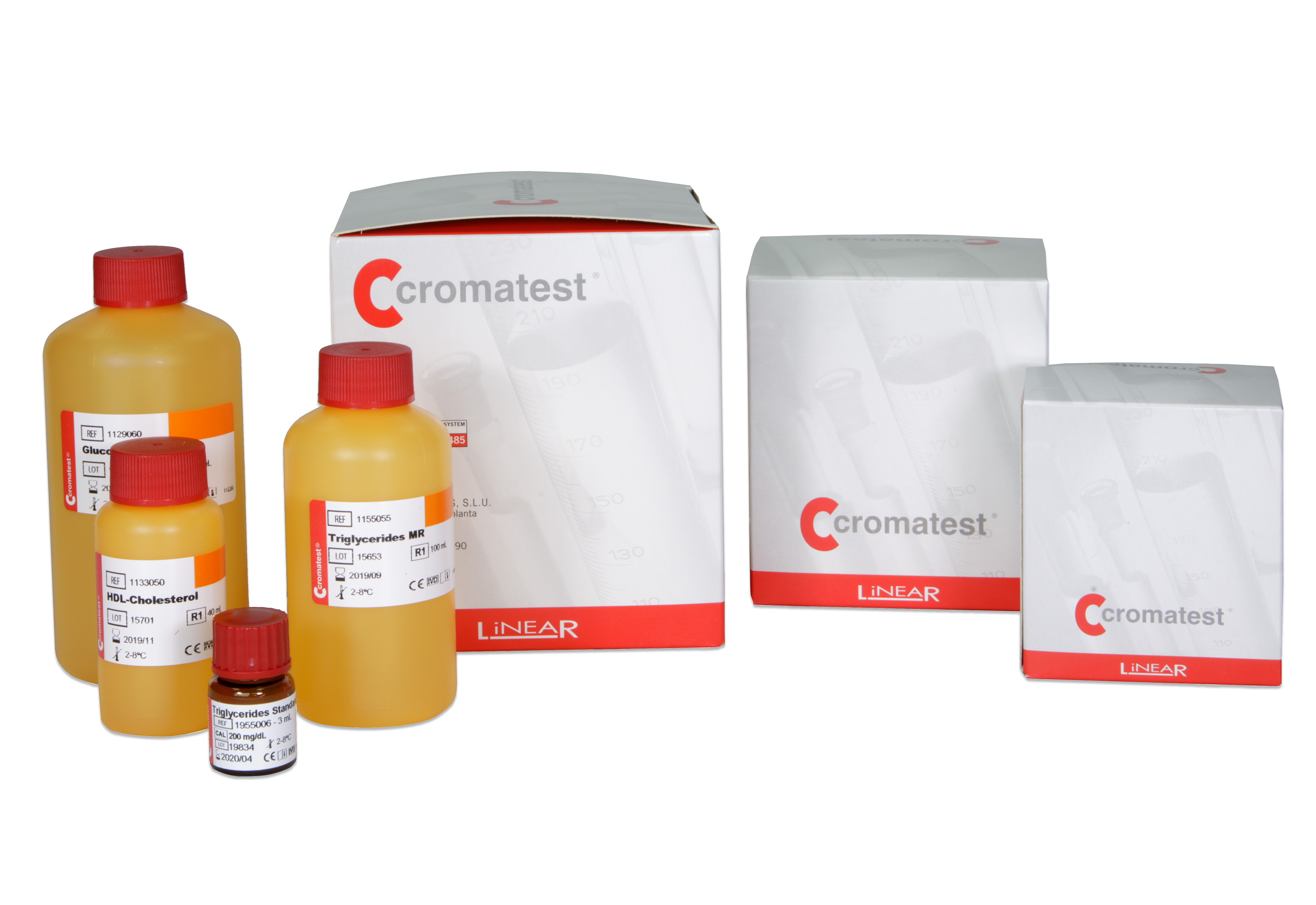

Cromatest
378 results
-

Amylase MR 1x20ml
1107049 -

Amylase MR 2x30ml
KR10060 -

Amylase MR 8x30ml
KR10062 -

Amylase MR 2x40ml
CT10060 -

AST/GOT BR opt. 3x100ml
1109010Aspartate aminotransferase (AST/GOT) catalyzes the transfer of the amino group from aspartate to alpha-ketoglutarate, forming glutamate and oxaloacetate. The latter is reduced to malate by malate dehydrogenase (MDH) in the presence of reduced nicotinamide adenine dinucleotide (NADH). The reaction is kinetically monitored at 340 nm by the decrease in absorbance resulting from the oxidation of NADH to NAD+, proportional to AST activity in the sample.
-

AST/GOT BR opt. 5x40ml
KR10072Aspartate aminotransferase (AST/GOT) catalyzes the transfer of the amino group from aspartate to alpha-ketoglutarate, forming glutamate and oxaloacetate. The latter is reduced to malate by malate dehydrogenase (MDH) in the presence of reduced nicotinamide adenine dinucleotide (NADH). The reaction is kinetically monitored at 340 nm by the decrease in absorbance resulting from the oxidation of NADH to NAD+, proportional to AST activity in the sample.
-

AST/GOT BR opt. 11x40ml
KR10075Aspartate aminotransferase (AST/GOT) catalyzes the transfer of the amino group from aspartate to alpha-ketoglutarate, forming glutamate and oxaloacetate. The latter is reduced to malate by malate dehydrogenase (MDH) in the presence of reduced nicotinamide adenine dinucleotide (NADH). The reaction is kinetically monitored at 340 nm by the decrease in absorbance resulting from the oxidation of NADH to NAD+, proportional to AST activity in the sample.
-

AST/GOT BR opt. 3x50ml
CT10072Aspartate aminotransferase (AST/GOT) catalyzes the transfer of the amino group from aspartate to alpha-ketoglutarate, forming glutamate and oxaloacetate. The latter is reduced to malate by malate dehydrogenase (MDH) in the presence of reduced nicotinamide adenine dinucleotide (NADH). The reaction is kinetically monitored at 340 nm by the decrease in absorbance resulting from the oxidation of NADH to NAD+, proportional to AST activity in the sample.
-

Acid Phosphatase 4x10ml
1100005The method is based on the hydrolysis of a-naphthyl phosphate at pH 5.0 by acid phosphatase (FAC), producing a-naphthol and inorganic phosphate. Pentanediol acts as a phosphate acceptor, increasing the reaction sensitivity. a-naphthol reacts with a diazonium salt, Fast Red TR*, forming a colored complex directly proportional to the FAC activity in the sample.
-

Albumin 2x50ml
1101000 -

Albumin 4x100ml
1101010 -

Albumin 4x250ml
1101025