
Clonatest
250 results
-

-

Iron Standard 100 µg/dL 1x3 ml
1938005 -

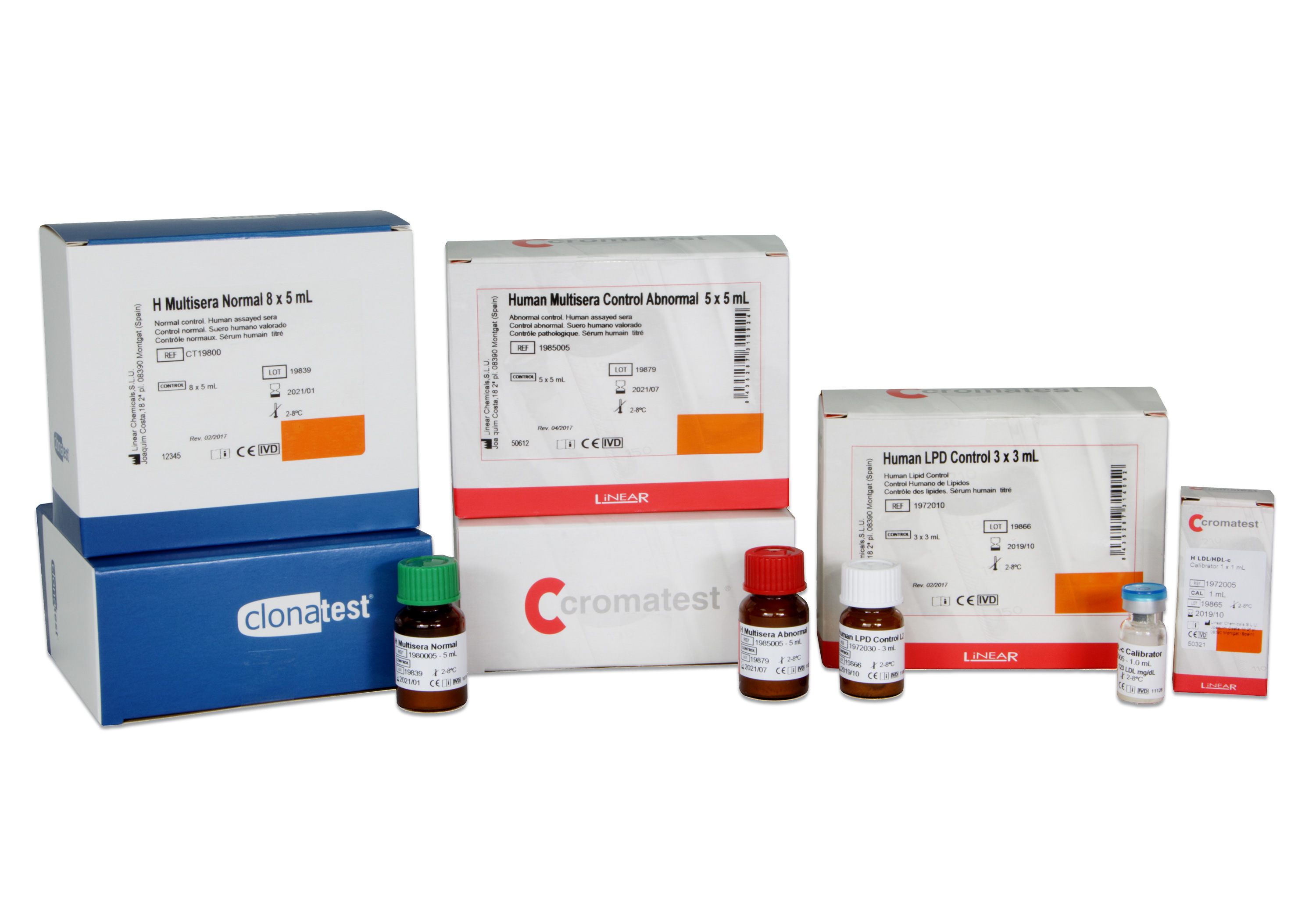
Human Multisera Normal 5x5 ml
1980005 -

Uric Acid MR 6x40 ml
CT10392Uric acid is oxidised by the action of uricase to allantoin and hydrogen peroxide. In the presence of peroxidase (POD) the mixture of dichlorophenol sulphonate (DCBS) and 4-aminoantipyrine (4-AA) are condensed by the action of hydrogen peroxide, forming a coloured quinonaimine proportional to the concentration of uric acid in the sample.
-

Urea/BUN BR 12x50 ml
CT10385Urea is hydrolysed by urease to ammonia and carbon dioxide. Ammonia is converted to glutamate by glutamate dehydrogenase (GlDH) in the presence of NADH and ketoglutarate. The reaction is measured kinetically at 340 nm through the decrease in absorbance resulting from the oxidation of NADH to NAD+, proportional to the concentration of urea present in the sample.
-

Uric Acid MR 2x50 ml
1161005Uric acid is oxidised by the action of uricase to allantoin and hydrogen peroxide. In the presence of peroxidase (POD) the mixture of dichlorophenol sulphonate (DCBS) and 4-aminoantipyrine (4-AA) are condensed by the action of hydrogen peroxide, forming a coloured quinonaimine proportional to the concentration of uric acid in the sample.
-

Uric Acid MR 10x60 ml
CT10395Uric acid is oxidised by the action of uricase to allantoin and hydrogen peroxide. In the presence of peroxidase (POD) the mixture of dichlorophenol sulphonate (DCBS) and 4-aminoantipyrine (4-AA) are condensed by the action of hydrogen peroxide, forming a coloured quinonaimine proportional to the concentration of uric acid in the sample.
-

-

Albumin Standard 5 g/dL 1x3 ml
1901005 -

Total Protein 6x40 ml
CT10352En la reacción del biuret se forma un quelato entre el ión Cu2+ y los enlaces peptídicos de las proteínas en medio alcalino con la formación de un complejo coloreado violeta cuya absorbancia se mide fotométricamente. La intensidad del color producido es proporcional a la concentración de proteínas en la muestra.
-

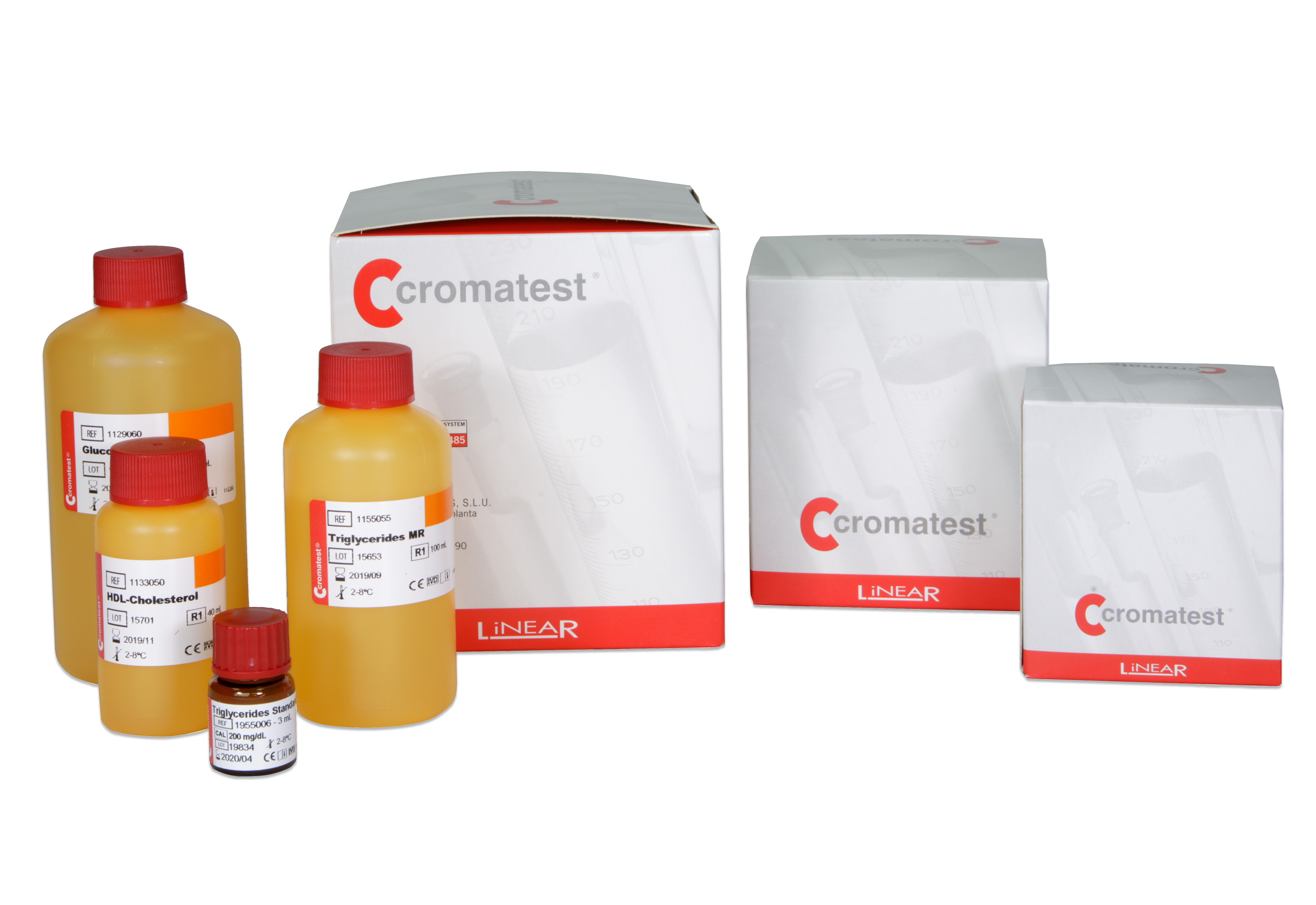
Triglycerides MR 2x50 ml
1155005The method is based on the enzymatic hydrolysis of serum triglycerides to glycerol and free fatty acids (FFA) by the action of lipoprotein lipase (LPL). Glycerol is phosphorylated by adenosine triphosphate (ATP) in the presence of glycerol kinase (GK) to form glycerol-3-phosphate (G-3-P) and adenosine diphosphate (ADP). G-3-P is oxidized by glycerophosphate oxidase (GPO) to dihydroxyacetone phosphate (DHAP) and hydrogen peroxide. In the presence of peroxidase (POD)
-

Total Protein 10x60 ml
CT10355En la reacción del biuret se forma un quelato entre el ión Cu2+ y los enlaces peptídicos de las proteínas en medio alcalino con la formación de un complejo coloreado violeta cuya absorbancia se mide fotométricamente. La intensidad del color producido es proporcional a la concentración de proteínas en la muestra.