
Clonatest
250 results
-

Creatine Kinase BR 3x50 ml
CT10162Creatine kinase (CK) catalyzes the reaction between creatine phosphate (CP) and adenosine 5'-diphosphate (ADP), forming creatine and adenosine 5'-triphosphate (ATP). The latter converts glucose to glucose-6-phosphate (G6P) in the presence of hexokinase (HK). G6P is oxidized to gluconate-6P in the presence of reduced nicotinamide adenine dinucleotide phosphate (NADP), catalyzed by glucose-6-phosphate dehydrogenase (G6PDH). The conversion is kinetically monitored at 340 nm through the increase in absorbance resulting from the reduction of NADP to NADPH, proportional to CK activity in the sample. The inclusion of N-acetylcysteine (NAC) in this method allows optimal enzyme activation.
-

Creatine Kinase MR 1x25 ml
1121005The higher activity of CK in normal sera is due to the isoenzymes CK-MM and CK-MB present in muscle and cardiac tissues. CK-BB is usually present at very low concentrations. Both creatine kinase (CK) enzymes are dimers formed by the association of two muscle (M) and brain (B) subunits. Immunoinhibition with a specific antibody for both MM subunits and the individual unit of CK-MB allows the determination of the B subunit. The activity corresponding to half of CK-MB is measured through the increase in absorbance resulting from coupled reactions.
-

Creatine Kinase MR 3x50 ml
CT10172The higher activity of CK in normal sera is due to the isoenzymes CK-MM and CK-MB present in muscle and cardiac tissues. CK-BB is usually present at very low concentrations. Both creatine kinase (CK) enzymes are dimers formed by the association of two muscle (M) and brain (B) subunits. Immunoinhibition with a specific antibody for both MM subunits and the individual unit of CK-MB allows the determination of the B subunit. The activity corresponding to half of CK-MB is measured through the increase in absorbance resulting from coupled reactions.
-

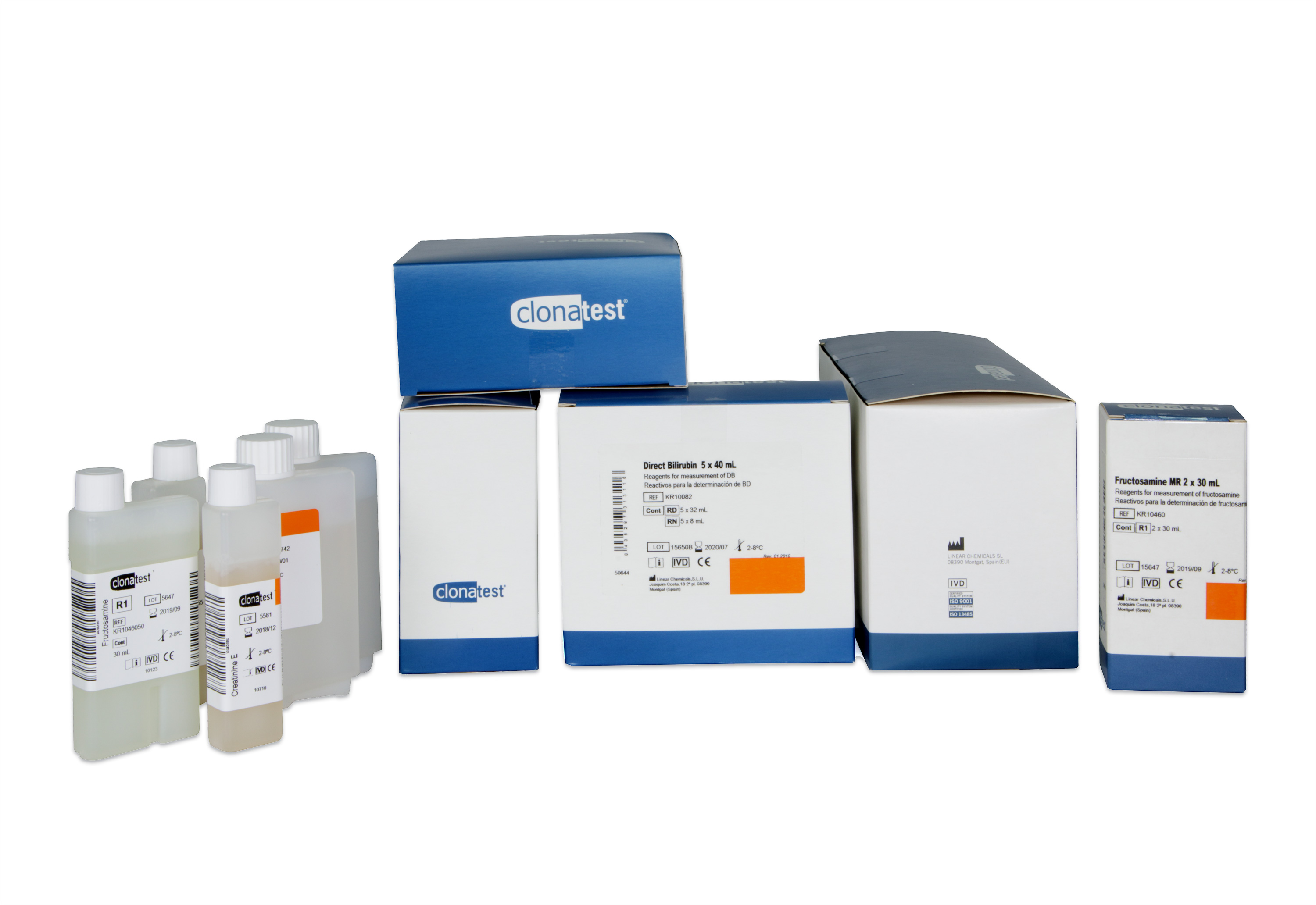
Fructosamine 2x30 ml
KR10460The assay is based on the reductive ability of ketoamines formed by non-enzymatic condensation between glucose and serum proteins (fructosamines) to reduce tetrazolium salts in alkaline solution to a photometrically measurable coloured complex. The oxidised tetrazolium salt, initiator of the reaction, is reduced by the action of fructosamine to form a purple formazan complex.
-

Creatinine Enzimatic 12x45 ml
CT10155This procedure is based on an improved enzymatic method originally designed to assess serum and urinary creatinine. The assay is performed in two stages. In the first stage, creatine is eliminated during the initial minutes of sample pre-incubation with creatinase. In the second stage, the addition of creatininase initiates the reaction, hydrolyzing creatinine in the sample in the presence of sarcosine oxidase (Sar OD) with the production of hydrogen peroxide:_x000D_ Creatinine + H2O → Creatine_x000D_ Sarcosine + H2O + O2 → H2O2 + Glycine + HCHO_x000D_ The hydrogen peroxide derived from the oxidase reaction is quantified by a Trinder-type reaction in which the chromogenic derivative HTIB and 4-aminoantipyrine (4-AA) condense in the presence of peroxidase (POD) to form a red quinonimine dye._x000D_ 4-AA + HTIB → Quinonimine + H2O_x000D_ The color development rate is proportional to the creatinine concentration in the sample.
-

-

Bilirubin Direct DPD 12x50ml
CT10085Direct (conjugated) bilirubin reacts with the diazonium salt 2,4-dichlorophenyldiazonium (2,4-DPD) in the presence of sulfamic acid, forming azobilirubin. This colored complex can be measured photometrically at 546 nm. Of the two fractions of bilirubin present in serum, bilirubin glucuronate (conjugated) and bilirubin free associated with albumin (unconjugated), only the first reacts directly, while the free bilirubin needs to be dissociated from the protein by an accelerator to react. Indirect bilirubin is calculated by the difference between total bilirubin (with accelerator) and direct bilirubin (without accelerator). The concepts of 'direct' and 'indirect' refer exclusively to the reaction characteristics in the presence or absence of accelerators or solubilizers and are only approximate equivalents to the two mentioned bilirubin fractions.
-

-

Calcium OCC 2x50ml
1115000 -

Cholinesterase 30 Test
1119005Cholinesterase (CHE) catalyzes the hydrolysis of the substrate butyrylthiocholine, forming butyrate and thiocholine. The latter reduces 5,5'-mercaptobis-2-nitrobenzoic acid (DMNB) to 5-mercapto-2-nitrobenzoate (5-MNBA), a colored compound. The reaction is kinetically monitored at 405 nm based on the rate of yellow color formation, proportional to CHE activity in the sample.
-

Cholesterol MR 2x50 ml
1118005This method for the determination of total cholesterol in serum is based on the use of three enzymes: cholesterol esterase (CE), cholesterol oxidase (CO), and peroxidase (POD). In the presence of the latter, the mixture of phenol and 4-aminoantipyrine (4-AA) condenses due to hydrogen peroxide, forming a colored quinonimine proportional to the concentration of cholesterol in the sample.
-

Cholesterol MR 6x40 ml
CT10142This method for the determination of total cholesterol in serum is based on the use of three enzymes: cholesterol esterase (CE), cholesterol oxidase (CO), and peroxidase (POD). In the presence of the latter, the mixture of phenol and 4-aminoantipyrine (4-AA) condenses due to hydrogen peroxide, forming a colored quinonimine proportional to the concentration of cholesterol in the sample.